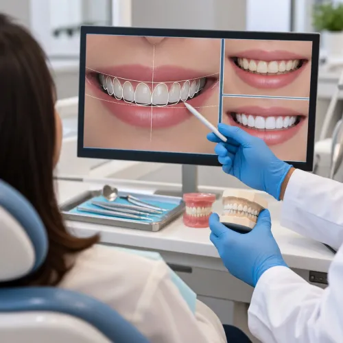
Deep periodontal pockets that remain after scaling and root planing can shelter calculus and inflamed tissue beyond the reach of routine instruments. By the end, you will understand how clinicians measure that problem, when flap surgery is appropriate, what happens during recovery, and which factors determine whether treatment remains stable.
Key takeaways
- Persistent deep pockets after scaling and root planing can justify flap surgery.
- Flap surgery exposes root surfaces so deposits and damaged tissue can be removed precisely.
- Expect soreness, swelling and modified brushing while the gum heals.
- Maintenance visits and daily plaque control help preserve surgical results.
What Makes Gum Disease Advanced Enough to Consider Flap Surgery?
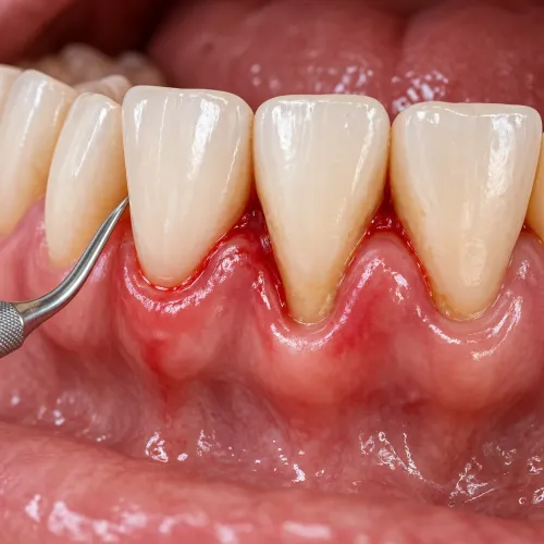
Advanced periodontitis is identified by persistent loss of gum attachment and supporting bone, not by redness alone. Periodontal pockets that remain deep after professional scaling and root planing indicate disease that closed instruments have not adequately controlled.
The assessment combines these findings:
- Periodontal probing depths in millimetres, recorded at several points around every tooth
- Bleeding on probing, which indicates inflamed tissue around the pocket
- Gum recession and the amount of remaining clinical attachment
- Tooth mobility and changes in bite or chewing comfort
- Dental X-rays showing the amount and pattern of bone loss
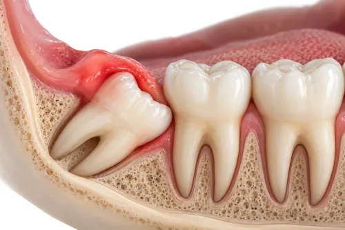
- Furcation involvement, meaning bone loss between the roots of a multirooted tooth
- Medical history, including diabetes control, smoking and medicines that affect healing
Flap surgery becomes relevant when residual pockets are deep, bleeding, or inaccessible after nonsurgical treatment and improved daily plaque removal. Surgical access lets the clinician see contaminated root surfaces, furcations and vertical bone defects that cannot be cleaned predictably through a closed pocket.
The decision also considers remaining bone support, root anatomy, whether the tooth can be restored, mobility and your ability to maintain plaque control. A gumline that looks healed does not prove the disease is controlled; follow-up probing, bleeding status and radiographic or clinical findings guide the decision.
Flap surgery does not automatically save every severely affected tooth.
Why Non-Surgical Treatment Comes First—and What Surgery Adds
Scaling and root planing is usually attempted before surgery because professional subgingival instrumentation can remove calculus and bacterial deposits from pocket surfaces without incisions. Improved daily plaque control then reduces the new deposits that keep inflammation active. If pockets remain deep or inaccessible after healing, surgery provides access that instruments cannot reliably achieve.
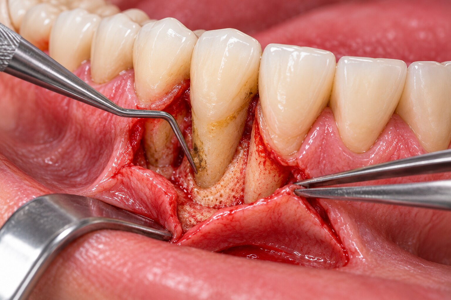
Surgery does not mean removing a strip of gum. Under local anaesthetic, the clinician makes gumline incisions and performs lifting the gum tissue to expose contaminated root surfaces and the pocket wall. Direct visibility changes cleaning from working through a narrow pocket to inspecting the entire root and underlying defect.
- Calculus and inflamed granulation tissue can be identified and removed under direct vision, including deposits hidden by pocket depth.
- Root surfaces can be instrumented more completely, while the pocket architecture can be assessed for areas that trap plaque.
- The gum can be repositioned against cleaned surfaces to create a shallower area that you can maintain at home.
The aim is better access, reduced pocket depth and improved daily cleaning, not a permanent cure for periodontitis. Bacteria will return if plaque control declines, so periodontal-maintenance visits and consistent home care remain necessary after either approach.
What Happens During Flap Surgery, and When Are Additional Procedures Used?
The clinician numbs the area, makes incisions along the gumline, and gently lifts a flap rather than removing a strip of gum. This exposes root surfaces, furcation areas and underlying bone defects, allowing direct access where instruments used through a closed pocket cannot clean predictably.
- Dental calculus and inflamed granulation tissue are removed from the pocket and exposed root surfaces.
- Contaminated root surfaces are carefully smoothed to remove bacterial deposits and create a cleaner surface for healing.
- Bone defects are cleaned and, when necessary, reshaped to reduce areas that trap plaque.
- The gum flap is repositioned around the teeth and secured with sutures.
Bone grafting is selected for specific intrabony defects where added material can support repair or fill part of the lost support. Guided tissue regeneration uses a barrier membrane, sometimes with graft material or a biologic agent, when the defect’s shape can protect the healing space. Neither approach regrows lost bone in every advanced case.
Systemic antibiotics are not routine after flap surgery. A clinician selects them only when the infection, medical risk, or treatment protocol provides a clear indication; unnecessary courses can cause adverse effects and promote antimicrobial resistance.
A furcation defect or deep vertical defect often determines whether added regenerative treatment is useful, while straightforward access cleaning may require no additional material.
What Can You Expect Before, During and After the Appointment?
Plan time for a review of your medical history, medicines and the number of teeth involved. Tell the clinician about diabetes, smoking, blood thinners and previous problems with anaesthesia; do not stop prescribed medicines without medical advice.
1. Local anaesthesia numbs the gum before the incision. You should feel pressure and movement, not sharp pain, while the gum is lifted, the roots are cleaned and the flap is sutured.
2. Expect postoperative swelling, light bleeding or oozing, soreness and an altered appearance. Bite on gauze for the instructed period, use an ice pack outside the cheek during the first day, and contact the clinic if bleeding does not settle with firm pressure.
3. Protect the sutures by avoiding brushing and flossing directly over the operated area until instructed. Use a chlorhexidine rinse exactly as directed; do not swish forcefully, spit hard, smoke or drink alcohol during the protected period.
4. Choose soft, lukewarm foods and chew on the opposite side. Take pain relief only as directed, and report worsening pain, swelling, fever or a persistent bad taste.
5. As inflammation resolves, gum recession can reveal more of the root. This tooth-root exposure may make teeth look longer and cause cold or touch sensitivity; desensitising treatment can help.
A review checks healing and removes non-dissolving sutures when appropriate. Recovery varies with the number of teeth treated and whether regenerative materials were placed.
How Are the Results Judged and Kept Stable Over Time?
A shallower pocket is not proof that every tooth is saved. Flap surgery improves access and reduces disease, but prognosis still depends on remaining bone support, root shape, tooth restorability and forces from the bite.
The factors that most influence a tooth’s outlook include:
- Bone loss pattern: an isolated, deep defect may be easier to manage than widespread support loss.
- Tooth mobility: increasing movement signals reduced support or excessive occlusal forces.
- Furcation involvement: bone loss between the roots of a molar makes cleaning and long-term maintenance harder.
- Plaque control, smoking, diabetes control and the patient’s ability to attend maintenance visits.
Follow-up evaluates healing through periodontal probing depths, bleeding on probing, plaque levels and tooth mobility. The clinician may compare dental X-rays with earlier images to assess bone changes, but appearance alone cannot confirm stability.
At Dr. Panjiars Dental Clinic, asking for these measurements helps you understand whether a treated tooth is healing or still needs risk management.
- Brush twice daily with a soft brush and clean between teeth using floss, interdental brushes or another prescribed aid.
- Stop smoking; tobacco is linked to poorer treatment response and recurrence.
- Manage blood glucose with your medical team; periodontal surgery cannot replace diabetes treatment.
- Attend periodontal-maintenance visits at the interval recommended for your risk.
Treated sites can relapse because surgery does not permanently remove the bacterial cause of periodontitis. Consistent daily plaque removal and professional monitoring keep the result stable.
Related service
Gum Health & Periodontal Flap Surgery Treats gum infections and deep pockets Restores healthy gum structure Prevents tooth loss and bone damage Promotes long-term oral hygiene and... View service → |
Frequently asked questions
What makes gum disease advanced enough to consider flap surgery?
Persistent attachment and bone loss, especially pockets that remain deep after scaling and root planing, can indicate that surgery deserves consideration.
Why does non-surgical treatment come before periodontal flap surgery?
Scaling and root planing reduce inflammation and remove deposits without surgery, while flap surgery adds direct access to deep roots and supporting tissues that closed instruments cannot adequately reach.
What happens during periodontal flap surgery?
The periodontist numbs the area, lifts the gum to access the root and bone, removes deposits and diseased tissue, then repositions and sutures the gum.
How are the results of flap surgery kept stable over time?
Results depend on reduced pocket depth, healthier attachment, controlled inflammation and stable bone support, followed by professional maintenance and consistent daily plaque removal.