Pain behind your second molar does not automatically mean the wisdom tooth must come out, but swelling, pus, fever or restricted mouth opening needs prompt assessment. By the end, you will know how impaction and pericoronitis differ, which warning signs require same-day care, what imaging can show and when monitoring or another treatment makes more sense than extraction.
Key takeaways
- Same-day assessment is needed for facial swelling, fever, difficulty swallowing, or limited mouth opening.
- Impacted teeth can trap food and cause pericoronitis around a partially erupted crown.
- Examination and dental X-rays distinguish wisdom-tooth problems from decay, gum disease, or jaw infection.
- Removal is not automatic; monitoring is reasonable when the tooth is symptom-free and disease-free.
How impaction and pericoronitis cause wisdom-tooth symptoms
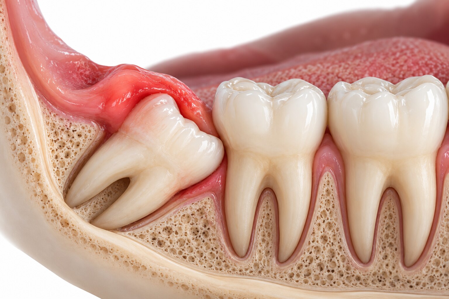
Impaction means a wisdom tooth cannot erupt into its normal position because bone, gum, or the neighbouring second molar blocks it. A partially erupted tooth has some crown visible; a fully impacted tooth remains beneath the gum or bone.
The direction of a wisdom tooth impaction describes its tilt:
- Mesioangular impaction: the crown leans forward, toward the second molar.
- Distoangular impaction: the crown leans backward, away from the second molar.
- Vertical impaction: the tooth is upright but lacks room to emerge.
- Horizontal impaction: the tooth lies on its side, with the crown directed toward the second molar.
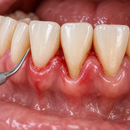
Brief tenderness, pressure, or gum soreness during eruption often settles without swelling or discharge. Pericoronitis is infection or inflammation around a partially erupted tooth: the gum flap over the crown traps food and bacteria, producing persistent or recurrent pain, swelling, foul taste, pus, bad breath, cheek biting, or difficulty opening your mouth.
Fever or trouble swallowing strengthens the concern for infection.
Pain behind the second molar can also come from decay, a crack, pulp inflammation, an abscess, or disease in that molar. One painful episode does not prove that extraction is needed, so arrange an examination if symptoms persist or recur.
Seek same-day care for facial or neck swelling, inability to swallow saliva, breathing difficulty, high fever, or severe restriction in mouth opening.
Which wisdom-tooth symptoms need same-day assessment
Pain behind the second molar, a swollen gum over wisdom tooth, foul taste or pus, or difficulty opening mouth and wisdom tooth pain all warrant prompt dental assessment. These signs often indicate pericoronitis, decay, an abscess, or disease affecting the neighbouring second molar.
A single painful episode does not prove that removal is necessary, but waiting can allow infection to worsen.
Arrange dental assessment soon if you have:
- Pain that persists, returns, or worsens behind the second molar
- Redness, tenderness, or swelling around a partially erupted tooth
- Foul taste, pus, bad breath, cheek biting, or food trapped under the gum flap
- Fever, tender neck glands, or trouble chewing and swallowing
Use this distinction to choose the right level of care:
| Sign | What it suggests | Action |
|---|---|---|
| Persistent pain or local gum swelling | Local inflammation, decay, or early abscess | Book a dental examination promptly |
| Fever with worsening pain, facial swelling, or reduced mouth opening | Infection extending beyond the tooth | Seek same-day dental care |
| Swelling spreading toward the eye or neck, inability to swallow saliva, breathing difficulty, high fever, or severe inability to open the mouth | Potentially dangerous spreading infection | Go to an emergency department immediately |
Do not wait for a routine extraction appointment when swelling is spreading or swallowing and breathing are affected. Antibiotics alone do not identify or remove the source; a dentist needs to examine the tooth and take appropriate radiographs.
How examination and imaging identify the real cause
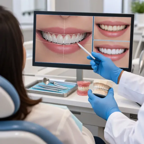
A dentist confirms whether the wisdom tooth is responsible by matching your pain’s location and triggers with clinical findings and dental images. Pain alone does not prove extraction is needed: decay, a crack, pulp inflammation, an abscess, or disease in the adjacent second molar can produce similar symptoms.
The wisdom tooth examination checks:
- The gum flap, trapped debris, swelling, pus and tenderness around the tooth
- Tooth mobility, decay, cracks and pain when biting or tapping
- The adjacent second molar for decay, bone loss or an abscess
- Mouth opening, jaw tenderness and signs that infection extends beyond the tooth
A panoramic radiograph is usually enough to show the tooth’s angulation, depth, root shape, neighbouring second molar and nearby bony structures. It commonly supports planning for an accessible tooth or a straightforward impaction.
Panoramic radiograph | Cone-beam CT wisdom tooth scan What it shows | A two-dimensional overview of teeth, roots and the inferior alveolar canal | Three-dimensional root, bone and canal relationships When it is enough | The tooth’s position is clear and nerve proximity does not appear difficult | The roots appear close to the inferior alveolar nerve or the view is unclear Why it is chosen | Lower radiation and adequate information for routine planning | More detail when it could change surgical technique, such as whether to consider coronectomy
A cone-beam CT scan is not automatically required for every extraction. The dentist recommends it when three-dimensional information can alter the plan or clarify surgical risk.
When removal is justified and when monitoring is reasonable
Extraction is justified when the wisdom tooth has disease, damages a neighbouring tooth, or repeatedly causes infection. Impaction alone, including a tooth that has never erupted, is not a sufficient reason.
| Finding | Usual decision | Why |
|---|---|---|
| Recurrent pericoronitis | Consider extraction | Repeated infection under the gum flap can return despite cleaning and medication. |
| Unrestorable decay in the wisdom tooth or second molar | Consider extraction | Caries that cannot be predictably restored leaves infection likely. |
| Abscess, cyst formation involving the wisdom tooth, or tumour | Remove the affected tooth or treat the lesion | Leaving the source permits infection, expansion or further bone damage. |
| Periodontal disease affecting the adjacent tooth | Consider extraction | Bone loss, caries or root resorption can threaten the functional second molar. |
| Disease-free, symptom-free impaction | Monitor | Routine removal offers no established general-health benefit and still carries surgical risk. |
Monitoring means scheduled examinations, appropriate radiographs and attention to whether the tooth remains cleanable, stable and free of decay, gum pockets, bone loss or cystic change. A previously quiet tooth needs reassessment if swelling, foul taste, gum bleeding or difficulty cleaning develops.
One painful episode does not prove that extraction is needed. Pain can come from a cracked tooth, pulp inflammation, decay in the second molar, an abscess or another dental condition; examination and radiographs must identify the source before treatment.
If the wisdom tooth is healthy and another tooth is responsible, treat that condition instead of removing a healthy tooth.
What to weigh before choosing wisdom-tooth surgery
The key decision is not simply whether the tooth hurts; it is how difficult and risky removal will be. Root curvature, tooth depth, angulation and bone coverage determine whether the tooth can be lifted out or needs bone removal and sectioning.
| Finding | Effect on surgery | Planning implication |
|---|---|---|
| Fully erupted, straight roots | Usually better access and easier delivery | May resemble removal of another molar |
| Deeply buried or horizontal tooth | More bone removal and a longer procedure | Expect greater swelling, jaw stiffness and recovery time |
| Curved, hooked or widely separated roots | Roots can fracture or resist removal | Ask how retained fragments would be managed |
| Roots close to the inferior alveolar canal | Raises concern for inferior alveolar nerve injury affecting lower-lip or chin sensation | Review panoramic imaging and consider cone-beam CT if it could change the approach |
A lower tooth that is very close to the nerve may be suitable for coronectomy: the crown is removed while the roots remain. It is not appropriate when the roots are infected or mobile, and retained roots can occasionally need later treatment.
Before proceeding, ask:
- What do the images show about root shape, depth, bone coverage and nerve distance?
- Would complete extraction or coronectomy give the safer result, and why?
- What should you expect during wisdom tooth extraction recovery, including pain, swelling, bruising and limited opening?
- How will you prevent or manage dry socket, infection, bleeding or altered sensation?
Smoking raises postoperative complication risk. At Dr. Panjiars Dental Clinic, ask the treating dentist to explain the imaging and alternatives in terms of your specific anatomy, not only the tooth’s position.
Frequently asked questions
How do impaction and pericoronitis cause wisdom-tooth symptoms?
An impacted tooth is blocked by bone, gum, or the second molar. Pericoronitis develops when bacteria and food collect under the gum flap over a partially erupted tooth, causing pain, swelling, and tenderness.
Which wisdom-tooth symptoms need same-day assessment?
Seek same-day dental assessment for facial swelling, fever, pus, difficulty swallowing or breathing, or restricted mouth opening.
How do examination and imaging identify the real cause?
A dentist checks the gum flap, tooth position, decay, bite, swelling, and mouth opening, then uses dental X-rays to assess impaction, infection, roots, bone, and the neighbouring molar.
When is wisdom-tooth removal justified?
Removal is justified when the tooth causes repeated infection, untreated decay, damage to the second molar, cyst-related changes, or persistent symptoms that treatment cannot predictably control.
When is monitoring a reasonable option?
Monitoring is reasonable when an impacted tooth has no symptoms, disease, or damage and can be reviewed through scheduled examinations and imaging.